ABOUT OUR
IMAGING SERVICES
MRI
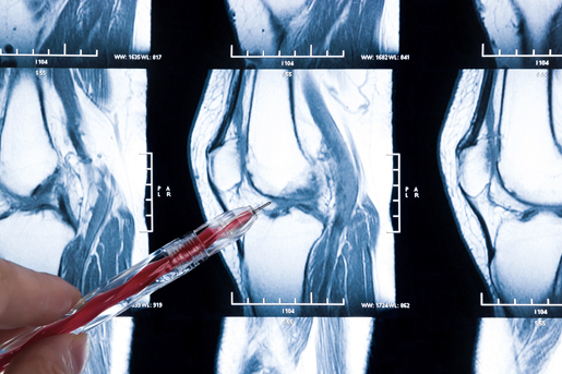
The human body is made up mostly of water. Simply put, magnetic resonance imaging or MRI works in concert with the water in your body to produce highly detailed pictures or “images” of your internal organs and structures. No radiation is used with this technology. MRI produces these images from any angle providing a wealth of diagnostic information quickly and easily. At Clearview MRI we offer high-field MRI scanners which set the standard in image quality. MRI is commonly used to examine the brain, hip, spine (cervical, thoracic, and lumbar), shoulder, knee, blood vessels, foot, and ankle. Prior to your exam, we’ll ask you to remove all metal – this includes hairpins, hearing aids, jewelry, snaps and zippers.
What should I expect during my MRI exam?


DIGITAL X-RAY
X-ray technology has been the baseline diagnostic tool in both hospitals and imaging centers for years. X-rays provide a quick and cost effective method for looking at bony structures in the body. Clearview MRI has chosen to provide Digital X-ray for the ease and convenience for our patients and their physicians. Digital X-ray eliminates the need to “process film” much the same way that your digital camera at home eliminates the need to have your film processed at your local drugstore. In addition to being very convenient, it also allows for a much better patient experience. No need to wait for film processing to make sure the image will provide the answer your doctor is looking for the image appears instantaneously.
What should I expect during my Digital X-Ray exam?
WOMEN’S IMAGING SERVICES PARTNERSHIP
Women everywhere have an instant bond when it comes to healthcare. They understand the pains of childbirth, the ups and downs of menopause, and they tell jokes about their mammogram experiences. While women are so great about laughing at all that life throws their way, these are often serious matters. Here at Clearview MRI, we want to help our patients by providing the highest quality women’s imaging in the most compassionate and comfortable environment possible.
A Breast MRI takes approximately 30-35 minutes to perform and requires that the patient receive an injection of a special contrast agent called gadolinium. It is very important that the patient be able to lie still during this test. You will be on your stomach carefully positioned on a special MRI coil for breast imaging. Breast MRI is a non-invasive procedure that doctors can use to see the inside of the breast without having to do surgery. Each exam produces hundreds of images of each breast for analysis. The technologist will go over your exam in detail before the exam begins. If you have any questions please feel free to call and speak with one of our technologists about what you can expect. (Please see general MRI section for additional details)
While ultrasound is still the initial test in imaging for either ovarian or uterine abnormalities, MRI is highly sensitive and accurate in evaluating the female pelvis for most disorders. Such as:
- Tumors of the ovaries, uterus or cervix. MRI is quite sensitive in detecting and characterizing a broad range of benign or malignant tumors of the female pelvis.
- PID (Pelvic Inflammatory Disease), Tubo-ovarian abscess or hydro salpinx
- Congenital abnormalities of the female reproductive system. Defining these anomalies has important implications in the treatment of certain kinds of female infertility.
- Uterine Fibroids or adenomyosis
- Ovarian cysts
- A variety of prolapse syndromes, including uterine or bladder prolapse, as well as vaginal and rectal prolapse. MRI can help in the diagnosis of complex prolapse syndromes. The initial diagnosis is made clinically, but many surgeons rely on MRI to distinguish between simple and complex prolapse syndromes prior to surgery.
A Pelvic MRI typically takes 30 -35 minutes. As with any MRI exam, patients will need to hold very still, they will go into the MRI scanner lying on their back, feet first. MRI contrast is typically not needed for these exams but may be required in some cases (Please see the general MRI section for additional details).


